 2017, Vol. 35
Institute of Oceanology, Chinese Academy of Sciences
Article Information
- GAO Qun(高群), HUANG Yong(黄勇)
- Oncholaimus zhangi sp. nov. (Oncholaimidae, Nematoda) from the intertidal zone of the East China Sea
- Chinese Journal of Oceanology and Limnology, 35(5): 1212-1217
- http://dx.doi.org/10.1007/s00343-017-6114-5
Article History
- Received Apr. 19, 2016
- accepted in principle May. 24, 2016
- accepted for publication Aug. 2, 2016


The biodiversity of free-living nematodes in the East China Sea was assessed using sediment samples. Sediment samples were collected from a number of sites from the intertidal to the sublittoral region during the past few years. Currently about 300 nematode species are known from these habitats (Yu et al., 2014; Jiang and Huang, 2015; Yu and Xu, 2015; Guo et al., 2016; Wang et al., 2016). During an investigation along the coast of the East China Sea in October 2014, a nematode of the genus Oncholaimus was collected from the sandy beach at Dongshan Island, Fujian Province and identified as a new species.
The genus Oncholaimus was described by Dujardin (1845). To date, 123 species in this genus have been recorded worldwide (Gerlach and Riemann, 1974: Chen and Guo, 2014: electronic database from Guilini et al., 2016), of which thirteen were synonyms of other species. Five species from the China Sea were described: Oncholaimus qingdaoensis Zhang and Platt (1983), O. sinensis Zhang and Platt (1983), O. multisetosus Huang and Zhang (2006), O. minor Chen and Guo (2014) and O. xiamenense Chen and Guo (2014). In this study a new species, O. zhangi sp. nov., from intertidal sediments of the East China Sea, is described.
2 METHODIn October 2014, sediment samples were collected from a coastal beach at Dongshan Island along the East China Sea. Sediment samples were collected using a syringe with a 2.6-cm internal diameter. The syringe was pushed into the sediment down to a depth of 8 cm. Samples were divided into two portions, representing the surface from 0–2 cm and deeper sediments from 2–8 cm, then fixed with 10% formalin in seawater. In the laboratory, samples were stained with 0.1% rose bengal for 24 h (Higgins and Thiel, 1988). The stained samples were washed with tap water to remove residual formalin and then sieved over two mesh sizes (500 and 42 μm) to separate macrofauna (500 μm) from meiofauna (42 μm). Heavier sediment particles were removed using centrifugation in Ludox-TM (50% colloidal silica, suspension in water; product of Sigma Aldrich Co., USA) with a specific gravity adjusted to 1.15 g/mL (de Jonge and Bouwman, 1977). Each sample was washed into a Petri dish with distilled water and the meiofaunawas sorted using a stereo microscope. Nematodes were transferred into 10 mL of 9׃1 (V׃V) solution of 50% ethanol׃pure glycerin in a cavity block. After the ethanol was slowly evaporated, the specimens were mounted in glycerin on permanent slides. A description of the new species was made from glycerin mounts using a differential interference contrast microscope (Nikon 80i). Line drawings were made with the aid of a camera lucida. All measurements are given in μm, and all curved structures were measured along the arc.
Type specimens (slide number: KMZ 0-2 (2) and paratype specimens (slide number KMZ 4-8-1 (1) and KMZ 3-1 (1) will be deposited in the Qingdao Institute of Oceanology, Chinese Academy of Sciences.
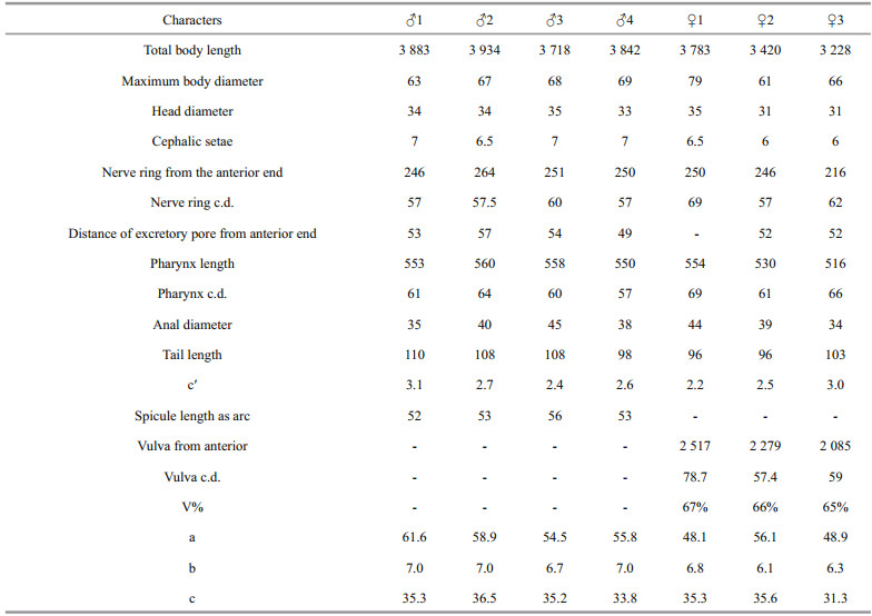
Abbreviations used in the figures and table (Figs. 1– 2, Table 1) are as follows: a: body length / max. bodydiameter; a.b.d.: anal body diameter; b: body length / pharynx length; c: body length / tail length; c.d.: corresponding body diameter; c': tail length / a.b.d; h.d.: head diameter; M: maximum body diameter; spic: spicule length along arc; V: corresponding body diameter of vulva; V%: position of vulva from anterior end expressed as a percentage of total body length.

|
| Figure 1 Oncholaimus zhangi sp. nov. a. lateral view of male anterior end; b. lateral view of female body; c. lateral view of the male tail end; d. lateral view of the male anterior end showing amphidial fovea; e. lateral view of the female anterior end; f. lateral view of spicule. |

|
| Figure 2 Oncholaimus zhangi sp. nov. a. lateral view of male anterior end; b. lateral view of female anterior end; c. lateral view of the male tail end; d. lateral view of the male tail end showing spicule. (Bars in a, b, c, d: 20 μm). |
|
Order ENOPLIDA
Family ONCHOLAIMIDAE Filipjev (1918)
Genus Oncholaimus Dujardin (1845)
Oncholaimus zhangi sp. nov. (Figs. 1–2, Table 1)
3.1 Type materialFour males and three females were collected in October 2014. Holotype, ♂1 on slide number KMZ 0-2 (2). Paratypes: ♂2 and ♂3 on slide number KMZ 4-8-1 (2), ♂4 and ♀1 on slide number KMZ 4-8-1 (1), ♀2 on slide number KMZ (0-2) (2) and ♀3 on slide number KMZ 3-1 (1).
3.2 Type locality and habitatSpecimens were collected from the surface intertidal sandy sediment from 0–2 cm and 2–8 cm depth at Dongshan Island, Fujian Province, in the East China Sea (23°43′22″N; 117°29′13″E).
3.3 EtymologyThis new species is named after Prof. ZHANG Zhinan in recognition of his work on nematodes.
3.4 MeasurementsAll measurement data are given in Table 1.
3.5 DescriptionMale: body cylindrical with blunt anterior end and tapered tail end. Cuticle is smooth with six longitudinal rows of stout somatic setae. Six rounded lips with six small and indistinct inner labial papillae. There are six outer labial setae and four cephalic setae set in a circle, almost equal in length. The large barrel-shaped buccal cavity is 33–35 μm deep and 18–20 μm wide, with heavily cuticularized walls and armed with three teeth. The left ventrosubventral tooth is larger than the right ventrosubventral and dorsal teeth. Amphidial fovea is shallow, cup-shaped, about 0.30–0.33 c.d and situated at the middle level of the buccal cavity (18– 20 μm from anterior end). The pharynx is cylindrical and muscular, 550–560 μm, surrounded by a nerve ring about mid-way along its length. Tail is conical, gradually tapering to cylindrical, and ventrally curved. A large ventral swelling is situated just posterior to the middle of the tail. A row of 6–7 ventral genital setae (5 μm) are present, three or four are anterior to the cloaca and three are posterior. In addition six pairs of short setae (4 μm) form a circular pattern around the cloaca. Two outstretched, opposed testes of about equal length, are both situated to the right of the intestine. Spicules are 1.2–1.5 a.b.d. long and slightly ventrally curved in the middle. The distal half is dilated with a pointed tip and the proximal end is slightly cephalated. Gubernaculum absent. Precloacal supplement absent. There are three caudal glands arranged in parallel, the anterior two extend beyond the level of the cloaca. The spinneret is conspicuous, appearing as a cap covering the tip of tail with a lateral terminal pore.
Female: similar to males except that somatic setae are absent and body size is slightly smaller compared with the male (female 3 228–3 783 μm vs male 3 718–3 934 μm). The reproductive system is monodelphic with a single anterior reflexed ovary, lying on the right side of the intestine. Vulva is located at 65%–67% of body length. The demanian system is a well developed tubular system located on the right side of the intestine. It consists of a long duct and a large uvette, the main duct with a terminal pore on the lateral side of the body. Spinneret is conspicuous as in a male.
3.6 Diagnosis and discussionThe genus Oncholaimus is characterized by a large buccal cavity with three teeth including a large left ventrosubventral tooth. The tail is short, straight or ventrally curved. Males are diorchic, with short spicules and no gubernaculum. Females are monodelphic with a single anterior ovary, and a demanian system (Platt and Warwick, 1983; Schmidt-Rhaesa, 2014). Oncholaimus zhangi sp. nov. is characterized by the male having a large ventromedian swelling just posterior to the middle of the tail, spicules are 52–56 μm in length, and circucloacal setae and ventral genital setae are present. The vulva is located at 65%–67% of the body length, and a similar conico-cylindrical tail is found in both males and females (no sexual dimorphism in tail shape). The genus Oncholaimus is a large genus. It is divided into two main groups according to the shape of the tail. In the Dujardini group, the tail is strongly bent ventrally (if it is not too short), stout, conical, rounded or sometimes provided with a distal cylindrical part. In the Paralangrunensis group, the tail is not (or only slightly) ventrally bent, slender, long and its greatest part is cylindrical (Wieser, 1953). Oncholaimus zhangi sp. nov. belongs to the Dujardini group. It most closely resembles O. sahariensis Coomans and Heyns (1983), O. balli Nicholas and Stewart (1984), O. cobbi Kreis (1932), O. longispiculosus Gerlach (1955), O. longus Wieser (1953) and O. oxyuris Ditlevsen (1911) in having a conspicuous ventral swelling on the rear-end of the tail in males. However, the new species can be distinguished from the above species using several characteristics including body size, cephalic setae length, spicule shape and length, no sexual dimorphism in tail shape and vulva position. Oncholaimus zhangi sp. nov. differs from O. sahariensis Coomans and Heyns (1983) by having a slightly larger body and longer spicules (3 234– 3 934 μm and 52 μm vs 2 720–3 270 μm and 47.5 μm), the presence of genital setae and tail shape is the same in both sexes. The tail is conico-cylindrical and ventrally arcuate in O. sahariensis males. However the tail of O. sahariensis females are broadly convex for about one anal body width, then abruptly narrows to a long peg of roughly the same length as the broad basal part. The new species differs from O. balli Nicholas and Stewart (1984) by having a larger body and longer spicules (3 234–3 934 μm and 52 μm vs 1 834–2 314 μm and 26–33.5 μm), and fewer circucloacal setae (6 pairs vs 16 pairs). Oncholaimus zhangi sp. nov. differs from O. cobbi Kreis (1932) by having a smaller body size (3 234–3 934 μm vs 4 523–5 117 μm), and as the vulva is placed in a more anterior position (V% 65%–67% vs 71%). In comparison with O. longispiculosus Gerlach (1955), O. zhangi sp. nov. have a longer body (3 234– 3 934 μm vs 2 239 μm), a longer tail (c' 2.2–3.1 vs c' 1.4–1.8) and shorter spicules (1.2–1.5 a.b.d. vs 3.2 a.b.d.). The new species has a longer body size (3 234–3 934 μm vs 2 970–3 025 μm), a wider cephalic diameter (33–35 μm vs 29 μm) and the vulva is located in a more anterior position (V% 65%–67% vs 73%) compared with O. oxyuris Ditlevsen (1911). The new species differs from O. longus Wieser (1953) by having a shorter body length (3 234–3 934 μm vs 5 000–7 630 μm), the excretory pore is in a more anterior position (1.5–1.7 h.d. vs 4 h.d. from the anterior end), the ventral swelling of the tail is more anterior (just posterior to the middle of the tail vs 3/4 from cloaca to tip of curved tail) and there are no precloacal papilla.
Oncholaimus zhangi sp. nov. also differs from other Oncholaimus species, O. qingdaoensis Zhang and Platt (1983) and O. xiamenense Chen and Guo (2014), described from the China Sea, which males have a single prominent ventral swelling at the tail. O. qingdaoensis Zhang and Platt (1983) has precloacal papilla in males, O. xiamenense Chen and Guo (2014) is characterized by sexual dimorphism in tail shape.
4 ACKNOWLEDGEMENTThe authors are very grateful to referees for their valuable comments and suggestions.
| Chen Y Z, Guo Y Q, 2014. Three new species of free-living marine nematodes from East China Sea. Zootaxa, 3841(1): 117–126. Doi: 10.11646/zootaxa.3841.1 |
| Coomans A, Heyns J, 1983. Oncholaimus sahariensis sp.n.(Nematoda) from the Algerian Sahara. Hydrobiologia, 107(3): 193–201. Doi: 10.1007/BF00036688 |
| de Jonge V N, Bouwman L A, 1977. A simple density separation technique for quantitative isolation of meiobenthos using the colloidal silica Ludox-TM. Marine Biology, 42(2): 143–148. Doi: 10.1007/BF00391564 |
| Ditlevsen H J, 1911. Danish freeliving nematodes. Vidensk.Meddr Dansk Naturh. Foren., 63: 213–256. |
| Dujardin F, 1845. Histoire Naturelle des HELMINTHES ou Vers Intestinaux. Libraire Encyclopédique de Roret, Paris: 652p. |
| Filipjev I N, 1918. Free Living Marine Nematodes of the Sevastopol Area. Trudy Osob Zoology Laboratory Sebastopol Biology Station, 2: 1–350. |
| Gerlach S A, Riemann F, 1974. The Bremerhaven checklist of aquatic nematodes Part 2. Veröffentlichungen des Instituts für Meeresforschung in Bremerhaven, 4: 405–736. |
| Gerlach S A, 1955. Zur Kenntnis der freilebenden marinen Nematoden von San Salvador. Z. Wiss. Zool., 158: 249–303. |
| Guilini K, Bezerra T N, Eisendle Flöckner U, Deprez T, Fonseca G, Holovachov O, Leduc D, Miljutin D, Moens T, Sharma J, Smol N, Tchesunov A, Mokievsky V, Vanaverbeke J, Vanreusel A, Venekey V, Vincx M. 2016. NeMys: world database of free-living marine nematodes. http://www.nemys.ugent.be. Accessed on 20160920. |
| Guo Y Q, Chen Y Z, Liu M D, 2016. Metadesmolaimus zhanggi sp. nov. (Nematoda:Xyalidae) from East China Sea, with a pictorial key to Metadesmolaimus species. Cahiers de Biologie Marine, 57(1): 73–79. |
| Higgins R P, Thiel H, 1988. Introduction to the Study of Meiofauna. Smithsonian Institution Press, Washington, DC: 488p. |
| Huang Y, Zhang Z N, 2006. New species of free-living marine nematodes from the Yellow Sea, China. Journal of the Marine Biological Association of the UK, 86(2): 271–281. Doi: 10.1017/S0025315406013129 |
| Jiang W J, Huang Y, 2015. Paragnomoxyala gen. nov.(Xyalidae, Monhysterida, Nematoda) from the East China Sea. Zootaxa, 4039(3): 467–474. Doi: 10.11646/zootaxa.4039.3 |
| Kreis H, 1932. Freilebende marine nematoden von den sundainseln Ⅱ. Oncholaiminae. (papers from Dr. Th.Mortensen's Pacific Expedition 1914-16, 61). Vidensk. Meddr Dansk Naturh. Foren., 93: 23–69. |
| Nicholas W, Stewart A, 1984. Oncholaimus balli n. sp.(Nematoda:Oncholaimidae) from a volcanic crater lake in Papua New Guinea, with observations on the colonisaion of the lake by nematodes. Nematologica, 30(1): 1–10. Doi: 10.1163/187529284X00419 |
| Platt H M, Warwick R M, 1983. Free-living Marine Nematodes. In:Kermack D M, Barnes R S K eds. Synopses of the British Fauna (New Series). Cambridge University Press, Cambridge: 274p. |
| Schmidt-Rhaesa A, 2014. Handbook of Zoology:Nematoda. Walter de Gruyter GmbH, Berlin: 759p. |
| Wang C M, Huang Y, 2016. Pseudolella major sp. nov.(Axonolaimidae, Nematoda) from the intertidal zone of the East China Sea. Chinese Journal of Oceanology and Limnology, 34(2): 295–300. Doi: 10.1007/s00343-015-4395-0 |
| Wieser W, 1953. Free-living marine nematodes I. Enoploidea.Acta Universltatis Lundensis (N. F. 2), 49(6): 1–155. |
| Yu T T, Huang Y, Xu K D, 2014. Two new species of the genus Linhystera (Xyalidae, Nematoda) from the East China Sea. Journal of the Marine Biological Association UK., 94(3): 515–520. Doi: 10.1017/S0025315413001811 |
| Yu T T, Xu K D, 2015. Two new nematodes, Pseudelzalia longiseta gen. nov., sp. nov. and Paramonohystera sinica sp. nov. (Monhysterida:Xyalidae), from sediment in the East China Sea. Journal of Natural History, 49: 509–526. Doi: 10.1080/00222933.2014.953224 |
| Zhang Z N, Platt H M, 1983. New species of marine nematodes from Qingdao, China. Bulletin of the British Museum Natural History (Zoology), 45(5): 253–261. |