 2018, Vol. 36
Institute of Oceanology, Chinese Academy of Sciences
Article Information
- YANG Jingwen(杨静文), XU Yuchao(徐玉超), XU Ke(许珂), PING Hongling(平洪领), SHI Huilai(史会来), LÜ Zhenming(吕振明), WU Changwen(吴常文), WANG Tianming(王天明)
- Molecular cloning and transcriptional analysis of a NPY receptor-like in common Chinese cuttlefish Sepiella japonica
- Chinese Journal of Oceanology and Limnology, 36(3): 892-904
- http://dx.doi.org/10.1007/s00343-018-6270-2
Article History
- Received Oct. 14, 2016
- accepted in principle Dec. 8, 2016
- accepted for publication Mar. 20, 2017


2 Marine Fisheries Research Institute of Zhejiang Province, Zhoushan 316022, China
Neuropeptides are small protein-like molecules used by neurons to transmit important regulatory signals in both vertebrates and invertebrates. Several neuropeptides are widely conserved in the animal kingdom from protostomes to deuterostomes (Blumenthal, 2010; Minakata, 2010; van Loy et al., 2010; Nässel and Wegener, 2011; Grimmelikhuijzen and Hauser, 2012). The neuropeptide Y (NPY) superfamily, including pancreatic polypeptide (PP) and peptide YY (PYY), is responsible for the central regulation of multiple physiological processes in vertebrates, such as food intake, energy balance, learning, and others (Redrobe et al., 1999; Beck, 2001; Hökfelt et al., 2008; Nguyen et al., 2011). The identification of its invertebrate counterpart showed that these NPY homologs have a characteristic C-terminus ending with an amidated Phe (F) rather than a Tyr (Y) residue and, hence are designated Neuropeptide F (NPF). The regulatory roles of NPFs in feeding, energy homeostasis, reproduction, and stress responses have been demonstrated in a variety of invertebrates including mollusks (Suzuki et al., 2002; Nässel and Wegener, 2011). Recently, NPF has been identified in numerous molluscan species such as the garden snail Helix aspersa (Leung et al., 1992), the marine mollusk Aplysia californica (Rajpara et al., 1992), the squid Loligo vulgaris (Smart et al., 1992), the gastropod snail Lottia gigantea (Veenstra, 2010), the pond snail Lymnaea stagnalis (de JongBrink et al., 2001), and so on. The signaling pathway activated by this neuropeptide is now becoming a new subject for studies to explore the functional regulatory mechanism.
The peptides of the NPY family display a wide array of biological activities that are thought to be mediated through a subfamily of G protein-coupled receptors (GPCRs) termed NPY receptors (Gerald et al., 1996). Five NPY receptors (NPYRs) have been identified from mammals to date: Y1, Y2, Y4, Y5, and Y6. In addition another receptor subtype (Y3) was suggested by pharmacological studies without supporting data from molecular cloning (Michel et al., 1998). Seven totally different NPY receptors have been described in vertebrates (Sundström et al., 2013). The evolution of NPYRs shows that vertebrate ancestors probably had three receptor subfamilies. The subfamily of Y1 includes the Y1, Y4, Y6, and Y8 receptors, the subfamily of Y2 is comprised of Y2 and Y7 (identified in zebrafish and frogs), and the subfamily of Y5 consists of only Y5, due to lack of close relatives of this receptor (Larhammar and Salaneck, 2004). In invertebrates, the NPYRs (also named NPFRs for their ligand NPFs) have been identified functionally for the insects Drosophila melanogaster, Bombyx mori, and Anopheles gambiae (Garczynski et al., 2002, 2005; Deng et al., 2014) and the mollusks L. stagnalis (Tensen et al., 1998) with additional predicted NPYRs from genomic sequences of Crassostrea gigas (Zhang et al., 2012) and Octopus bimaculoides (Albertin et al., 2015). With limited exploration of the NPYRs, the mechanisms by which NPY/NPYRs regulate multiple physiological activities in mollusks have not been discovered.
The cuttlefish Sepiella japonica (Phylum: Mollusca, Class: Cephalopoda) is distributed mainly in coastal regions of Zhejiang and Fujian provinces, and is now becoming an important aquatic species. Recently, several studies have focused on the reproduction and developmental regulation of S. japonica (Furuya, 2008; Cao et al., 2016; Lü et al., 2016a, b; Yan et al., 2016). Here, we describe the cDNA cloning of a S. japonica NPYR-like (SjNPYR-like) that is closely related in sequence to L. stagnalis NPFR and the vertebrate NPY receptor type 2. Further bioinformatic analysis was conducted, and cellular location in vitro and transcriptional detection of SjNPYR-like in different tissues and developmental stages were investigated. The results suggest that SjNPYR-like is phylogenetically close to the Y2 family, has the typical characteristics of a transmembrane GPCR, and mediates physiological functions in feeding and energy homeostasis during development.
2 MATERIAL AND METHOD 2.1 Sample collectionCommon Chinese cuttlefish S. japonica was collected from the culture pool at aquaculture station of the Marine Fisheries Research Institute of Zhejiang on the island of Xishan, Zhoushan, Zhejiang Province, China. To evaluate the expression profile of SjNPYRlike in different tissues and at different stages during the female reproductive cycle, ovaries were collected at four gonadal development stages categorized using a histological method. The different gonadal development stages (period Ⅰ—oogonium production period, period Ⅲ—interstitial growth period, period Ⅱ—protoplasmic growth period, and period Ⅳ trophoplasmic growth period) are defined following the published article (Yan et al., 2016). Different tissues such as brain, liver, gill, branchial heart, and muscle of females in period Ⅱ, as well as brain, liver, and ovary of females in all four stages (periods Ⅰ to Ⅳ) were collected for further gene expression analysis. All collected tissues were immediately frozen and stored in liquid nitrogen until RNA extraction was performed.
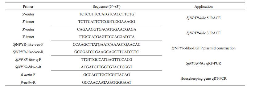
2.2 RNA extraction and cDNA synthesis using rapid amplification of cDNA ends (RACE)The total RNA for the 5′ RACE assay was extracted from the liver of S. japonica. RNA was extracted using Trizol reagent (TaKaRa, Japan) and phenol/ chloroform. 10.0 mg total RNA were used to perform the 5′ RACE protocol and 1.0 mg was used for the 3′ RACE protocol using FirstChoice® RLM-RACE Kit (Life Technologies, Madison, WI, USA) following the manufacturer's instructions. The cDNA products for the next PCR steps were stored at -20℃. The specific primers used for the RACE assays are listed in Table 1.
|
Comparison of the cDNA sequences with known sequences published in GenBank to determine percent identity was computered by online BLAST tools (BLASTX 2.5.1+; http://blast.ncbi.nlm.nih.gov). The encoding sequence of the SjNPYR-like was translated into amino acid sequence by BioEdit 7.0.5.3. The N-glycosylation sites were predicted using the NetNGlyc 1.0 Server (http://www.cbs.dtu.dk/services/NetNGlyc/) and the phosphorylation sites were deduced by NetPhos 2.0 Server (http://www.cbs.dtu.dk/services/NetPhos/). The physicochemical properties of these proteins were predicted using ProtParam (http://www.expasy.org/tools/protparam. html). The predicted amino acid sequence of the SjNPYR-like and other homologs were aligned using online Pairwise Sequence Alignment tool (http://www.ebi.ac.uk/Tools/psa/). The transmembrane region of SjNPYR-like was predicted using TMpred (http://www.ch.embnet.org/software/TMPRED_form.html). The prediction of protein domains were performed using online SMART (http://smart.emblheidelberg.de/) and InterProScan (http://www.ebi.ac.uk/InterProScan/). The NPYR 3D structure was modeled using SWISS-MODEL (http://swissmodel.expasy.org/). Analysis of the protein secondary structure was done using PredictProtein (http://www.predictprotein.org/). Based on the Maximum Likelihood (ML) Statistical Method, the phylogenetic tree was constructed using MEGA 5.1.
2.4 Mammalian expression vectors constructionTo amplify the full-length encoding sequence of SjNPYR-like, primers were designed based on its full-length cDNA sequence and to allow for subcloning into the pEGFP-N1 plasmid (Table 1). The PCR products were inserted into the final pEGFP-N1 expression vector using the HidⅢ and KpnI restriction enzymes (Beyotime, Haimen, China) and Rapid DNA Ligation Kit (Beyotime, Haimen, China). The constructed vector was named SjNPYRlike-EGFP, and sequenced to verify the correct reading frame.
2.5 Transfection and confocal microscopyThe HEK293 cells (human embryonic kidney cell line) was cultured in equilibrated growth medium (Dulbecco's modified Eagle's medium (DMEM), 10% fetal bovine serum (FBS, HyClone, Logan, UT, USA), 4 mmol/L-glutamine (Invitrogen, Madison, WI, USA)) at 37℃ in a humidified atmosphere containing 5% CO2. SjNPYR-like-EGFP was transfected into HEK293 cells using with X-tremeGENE HP DNA transfection reagent (Roche Applied Science, Indianapolis, USA) following to the manufacturer's instructions. After 12–16 h, the transfected HEK293 cells were seeded onto glass coverslips. After 16–24 h, cells were stained using DiI as the cell membrane probe (Beyotime, Haimen, China) for 5–10 min, then fixed with 4% paraformaldehyde at room temperature for 15 min, and finally stained by DAPI (Beyotime, Haimen, China) for 10 min incubating.
2.6 Real-time quantitative PCR (qRT-PCR)Total RNA from brain, liver, ovary, gill, branchial heart, and muscle tissues was extracted from female cuttlefishes in late vitellogenic stage. Moreover, brain, liver and ovary were collected at four gonadal development stages. Reverse transcription was conducted using the M-MLV reverse transcriptase(TaKaRa, Japan) and products was kept at -20℃ for the qRT-PCR analysis. β-actin was chosen as the internal control (housekeeping) gene. Specific primer sequences for β-actin were from previous reports (He et al., 2014; Yan et al., 2016). qRT-PCR primers specific for SjNPYR-like (Table 1) were designed according to the SjNPYR-like CDS sequence. The qRT-PCR assays were carried out by the ABI 7500 Software v2.0.6 (Applied Biosystems) and performed using the SYBR PrimeScript™ RT reagent Kit (TaKaRa, Japan).
The relative transcriptional level of SjNPYR-like was calculated using the 2-ΔΔCt method (Livak and Schmittgen, 2001). All data are presented as mean±S.D. (standard deviation). Differences were tested using PASW Statistics 18.00 (SPSS Inc., Chicago, IL, USA) with one-way Analysis of Variance (ANOVA) followed by Tukey's post hoc test. The significance level was set at 0.05.
3 RESULT 3.1 Isolation and characterization of SjNPYR-like cDNAThe full-length cDNA sequence of SjNPYR-like, 1 902 bp long, was cloned from S. japonica (GenBank accession No. KX683395). The sequence contained an open reading frame (ORF) of 1 182 bp encoding a protein of 393 amino acids, with 492 bp of 5′ untranslated region (5′ UTR), and 228 bp of 3′ UTR (Fig. 1). The predicted protein had a theoretical molecular weight of 45.54 kDa and an isoelectric point (pI) of 8.13. The putative SjNPYR-like was classified as belonging to the class A GPCR family according to the predicted 7TM_1 domain in amino acid sequence of this receptor. Meanwhile, the SjNPYR-like amino acid sequence contained several potential sites for modification (Fig. 1). Five potential N-linked glycosylation sites were present, with three in the extracellular N-terminal domain (at amino acids 2, 10, and 14) and two in the intracellular C-terminal domain (aa 339 and 365). Two conserved cysteine residues were present at positions 105 and 182 within the EC1 and EC2 domains. Twelve potential phosphorylation sites were present at five Serine residues (aa 115, 231, 234, 236, and 349), two Threonine residues (aa 96 and 379), and five Tyrosine residues (aa 15, 180, 309, 343, and 374).

|
| Figure 1 Full-length cDNA and deduced amino acid sequences of SjNPYR-like The predicted 7TM_1 conserved domain is shaded in grey. The conserved transmembrane domain regions (TM1-TM7) are noted with black underline. The putative N-glycosylated sites and phosphorylation sites are marked with black triangles and boxed by full lines, separatively. The termination codon is marked with an asterisk. |
The deduced amino acid sequence of S. japonica NPYR-like was compared with those of nine other homologous NPYR sequences (NPFRs, NPY2Rs, and NPY7Rs) that varied in length from 336 to 481 amino acid residues. Pairwise ClustalW analysis of amino acid sequences was carried out to evaluate homology relationships. The predicted S. japonica NPYR-like amino acid sequence showed high identity to the predicted NPY2R sequence of Octopus bimaculoides (81%), and other functionally identified NPFR and NPYR amino acid sequences, albeit with lower identity (ranging from 29% to 39%). Multiple sequence alignment analysis revealed conservation in the seven transmembrane (7TM) domains of NPYR sequences from various species including S. japonica (Fig. 2). Apart from S. japonica NPYR and O. bimaculoides NPY2R, significant variation was seen in the lengths of the N terminal, EC2, IC3, and C terminal regions (Fig. 2). The protein structure of SjNPYR-like was predicted using SWISS-MODEL (Fig. 3a) while secondary structure was predicted by PredictProtein (Fig. 3b). Homology modeling revealed that this protein was similar to human OX2 orexin receptor (4s0v.1.A) in the Protein Data Bank. The protein binding regions were predicted and marked on the constructed model.

|
| Figure 2 Alignment of the deduced SjNPYR-like amino acid sequence with sequences of other species Sequences of Octopus bimaculoides NPY2R (ObNPY2R), Lymnaea stagnalis NPFR (LsNPFR), Drosophila melanogaster NPFR (DmNPFR), Anopheles gambiae (AgNPFR), Bombyx mori NPFR (BmNPFR), Homo sapiens NPY2R (HsNPY2R), Epinephelus coioides NPY2R (EcNPY2R), Takifugu rubripes NPY7R (TrNPY7R), and Callorhinchus milii NPY7R (CmNPY7R) were obtained from GenBank database (Suppl. Table 1). The mutiple sequences alignment was prformed using online Pairwise ClustalW. The conserved transmembrane domain regions (TM1-TM7) are noted with black lines above. The extracellular and intracellular domains (EC, IC) are marked above. The black triangles indicate predicted phosphorylation sites and black spots indicate predicted N-linked glycosylation sites. |

|
| Figure 3 Predicted SjNPYR-like protein structure and domain organization a. predicted 3D structure of the SjNPYR-like protein. The conserved transmembrane domain regions (TM1-TM7), three extracellular and intracellular domains (EC, IC) are noted. The putative 3D structure of the SjNPYR-like protein was modelled by online SWISS-MODEL server; b. the conserved transmembrane regions and protein binding regions of SjNPYR-like. The dashboard overview was generated by online PredictProtein server. |
To examine the relationship of SjNPYR-like with NPYRs from various other species, neuropeptide FF receptors (NPFFRs) and Pyroglutamylated RFamide peptide receptors (QRFPRs), a phylogenetic tree was constructed with Mega 5.1 using the ClustalW multiple alignment and the protein sequences of SjNPYR-like and 45 alternate NPYRs, 6 NPFFRs and 6 QRFPRs from the gene bank (Fig. 4). Vertebrate NPYRs are categorized into three major clusters corresponding to the Y1 subfamily (NPY1R, NPY4R, and NPY6R), the Y5 subfamily (NPY5R), and the Y2 subfamily (NPY2R and NPY7R). Invertebrate NPYRs were separated into two distinct groups and were relatively close to the vertebrate Y2 subfamily. NPFFRs and QRFPRs clustered together into a seperated group. The deduced SjNPYR-like protein sequence grouped with the O. bimaculoides NPY2R and formed a cluster with the functionally identified NPFRs from D. melanogaster, B. mori, and A. gambiae. Meanwhile, the L.stagnlis NPFR and predicated C. gigas NPYR formed the second group of invertebrate NPYRs.

|
| Figure 4 Phylogenetic tree of SjNPYR-like amino acid sequences with 45 related NPYR homologs, 6 NPFFRs and 6 QRFPRs The tree was generated by MEGA 5.1 software based on Maximum Likelihood (ML) algorithms with 1 000 bootstrap replications. Y1R: neuropeptide Y1 receptor; Y2R: Y2 receptor; Y4R: Y4 receptor; Y5R: Y5 receptor; Y6R: Y6 receptor; Y7R: Y7 receptor; Y1: Y1 subfamily; Y2: Y2 subfamily; Y5: Y5 subfamily; NPFR: neuropeptide F receptor; NPFFR: neuropeptide FF receptor; QRFPR: Pyroglutamylated RFamide peptide receptor. * indicates the invertebrate NPYRs (NPFRs) which have been identified by functional criteria. Accession numbers are listed in Suppl. Table 2. |
To confirm the cellular membrane location of SjNPYR-like, the enhanced green fluorescent protein (EGFP) was fused to the C terminus of the receptor, and the resulting construct was expressed in human embryonic kidney 293 (HEK293) cells. Significant expression of this recpetor on cell surface was observed under confocal microscopy (Fig. 5). Result suggests the location of SjNPYR-like in the cell membrane in HEK293 as a transmembrane receptor, and the C-terminal EGFP tag does not affect SjNPYRlike expression.

|
| Figure 5 Confocal microscopy of HEK293 cells expressing the SjNPYR-like-EGFP fusion protein Transiently expressing SjNPYR-like-EGFP cells were stained with cell membrane probe (DiI) and cell nucleus probe (DAPI), and detected by confocal microscopy. All images are representative of at least three independent experiments. |
Different tissues of S. japonica were sampled at four stages during the gonadal development process, and body mass and ovarian wet weight were recorded (Fig. 6). From relative quantification analysis (Fig. 7), SjNPYR-like expression was ubiquitous in multiple tissues during the interstitial growth period (Ⅲ). SjNPYR-like expression was high in the brain, liver, and ovary, but low in the gill, branchial heart, and muscle. To determine the expression profile of SjNPYR-like in the brain, liver, and ovary tissues of S. japonica during different gonadal development stages, the SjNPYR-like mRNA level was determined (Fig. 8). In all three tissues, the transcriptional level of SjNPYR-like was relatively high during the protoplasmic growth period (Ⅱ) and the interstitial growth period (Ⅲ), and then decreased to low levels in the trophoplasmic growth period (Ⅳ), with an extremely low gene expression level in the ovary during the trophoplasmic growth period (Ⅳ).

|
| Figure 6 The body mass and ovarian wet weight during gonadal development Tissues were collected from females in different gonadal development stages: (Ⅰ) oogonium production period, (Ⅱ) protoplasmic growth period, (Ⅲ) interstitial growth period, and (Ⅳ) trophoplasmic growth period. Values indicate mean±S.D. n=6. |

|
| Figure 7 Relative expression of SjNPYR-like in different tissues of females in period Ⅱ (protoplasmic growth period) Total RNA was isolated and purified from brain, liver, ovary, gill, branchial heart, and muscle. The relative expression value was normalized against the expression of β-actin (the housekeeping gene). Different lowercase letters noted above the value bars indicate the significant differences between different tissues (P < 0.05). |

|
| Figure 8 Relative expression of SjNPYR-like in brain, liver, and ovary tissues from females in different gonadal development stages Ⅰ: oogonium production period; Ⅱ: protoplasmic growth period; Ⅲ: interstitial growth period; Ⅳ: trophoplasmic growth period. The relative expression value was normalized against the expression of β-actin (the housekeeping gene). Different lowercase letters above the value bars indicate the significant differences between different tissues (P < 0.05). |
The common cuttlefish S. japonica, distributed mainly in the coastal regions of Zhejiang and Fujian provinces, is an important aquatic species harvested in China (Wu and Tang, 1990; Wu et al., 2010). To improve production, artificial breeding methods have been developed, and these aquaculture techniques have been applied successfully in China. However, precocious puberty of cultured S. japonica became apparent, and weakened the development of this industry along the coast of China. A good understanding of the regulation of growth and reproduction in this species has become increasingly important. Recent studies on the control of growth and reproduction in cephalopods suggest that NPYR and its ligand NPY play important roles in feeding behavior and regulation of sexual maturation via central neuroendocrine control (Di Cristo, 2013). However, there are no publications on the regulatory roles of the NPY/NPYR system in S. japonica.
This study reports for the first time cloning of the NPYR homolog of S. japonica. The NPYR cDNA sequence contained 393 amino acid residues (Fig. 1). The predicted conserved protein domain 7TM_1 of this putative amino acid sequence indicated SjNPYRlike belongs to the class A (rhodopsin) GPCR family (Marchler-Bauer et al., 2015). In addition, five N-linked glycosylation sites and 12 phosphorylation sites were predicted. These putative sites are suggested mainly related with the protein activity, function, diversity, localization, cell signaling, and transcriptional modification (Arnold et al., 2007; Miedlich and Abou-Samra, 2008). Further multiple sequence alignment analysis indicated that SjNPYRlike was conserved with other invertebrate NPYRs and vertebrate Y2 receptors, especially in the transmembrane regions (Fig. 2). Otherwise, lower identity in the C-terminal and N-terminal regions of NPYRs from different species could be related to the diverse ligands and signal transduction properties associated with the different receptors. The high conservation between SjNPYR-like and putative ObNPY2R of the entire protein sequences, N-linked glycosylation sites, and phosphorylation sites suggested these two receptors belong to the same subtype.
NPYRs mediate cell signal transduction through binding to the agonist NPYs and activating the coupled G protein. Significant progress in research on GPCRs, the ligand-binding modes, receptor activation, and regulation of subsequent internalization mechanisms has been made by many organizations. The intracellular loops and C-terminus are involved in G protein-mediated signaling, internalization, and phosphorylation (Duvernay et al., 2004; Kristiansen, 2004). The extracellular loops and N-terminus are probably necessary for ligand recognition and binding (Merten et al., 2007). In this study, bioinformatics analysis was used to model the SjNPYR-like 3D structure and predict its secondary structure. Since the NPYR crystal structure has not been determined, homology of the predicted SjNPYR-like model with the 4s0v.1.A structure (human OX2 orexin receptor) from the Protein Data Bank was analyzed (Fig. 3a). SjNPYR-like protein binding regions were analyzed, and potential sites that may be involved with ligand binding were predicted: two (aa 175–177 and 187–190) in the EC2 domain, and one (aa 282–283) in the EC3 domain. Furthermore, regions that are thought to play key roles in G protein coupling were predicted (Fig. 4b): four sites (aa 223, 234–236, 239–240, and 245) in the IC2 domain, and one site (aa 329) in the intracellular C-terminal tail.
Phylogenetic analysis was carried out to investigate the evolutionary relationships between SjNPYR-like and NPYRs from vertebrates and invertebrates, as well as 6 NPFFR sequences and 6 QRFPR seuqences were also included in this phylogenetic tree to see if the cuttlefish sequences clearly belongs to the NPYR clade to the exclusion of other receptor families (Fig. 4). Vertebrate NPYRs clustered into three major groups corresponding to the Y1, Y2, and Y5 subfamilies. Invertebrate NPYRs were separated into two distinct groups and were relatively close to the vertebrate Y2 subfamily. The NPFFRs and QRFPRs clustered together with two seperated groups. However, further studies on the L. stagnalis NPYR (NPFR) suggested that this invertebrate NPYR resembles the Y1 subtype more than the Y2 subtype (Tensen et al., 1998). In addition, the Drosophila melanogaster NPYR (NPFR) was suggested to be not closely related to the Y2 subtype (Garczynski et al., 2002). This indicates experimental data on the signaling pathway characteristics of invertebrate NPYRs is crucial for subtype classification. The SjNPYR-like clustered with the putative NPY2R of O. bimaculoides with a high sequence identity of 81%, indicating that SjNPYR-like was closely related to ObNPY2R phylogenetically and evolutionarily, consistent with the evolutionary relationship of these two species. And it also clustered with the functionally identified NPFR of D. melanogaster, B. mori, and A. gambiae, suggesting its functional relationship with NPFRs.
GPCRs are described as seven-transmembrane receptors because they pass through the cell membrane seven times, and are located in the plasma membrane to sense molecules outside the cell and activate signal transduction pathways inside (Trzaskowski et al., 2012). The cell surface localization property of SjNPYR-like is crucial for its functional activity. To further assess the characteristics of SjNPYR-like as a transmembrane receptor, EGFP was fused to the C-terminus of SjNPYR-like and expressed in HEK293 cells. The expression of this recptor on cell surface was demonstrated by confocal microscopy (Fig. 5). With the putative 7TM domain structure of SjNPYRlike from structure modeling (Fig. 3), this result suggested that the cell surface localization property of SjNPYR-like was consistent with functioning as a transmembrane G protein-coupled receptor. The interaction between this receptor and its ligand should be investigated in future to clarify the signaling pathway and the details of the cellular responses.
NPYRs are distributed ubiquitously in the central nervous system and peripheral tissues to mediate diverse physiological functions (Silva et al., 2005; Hausman et al., 2008; Pezeshki et al., 2012; Shi et al., 2012). The gene expression of SjNPYR-like in various tissues was demonstrated here, and qRT-PCR analysis indicated that SjNPYR-like mRNA was detected at high levels in tissues that are known to be involved in developmental and reproductive processes, such as the brain, liver, and ovary (Fig. 7). NPY is known to play a key role in promoting food intake and energy budgeting in vertebrates (Frankish et al., 1995; White and Martin, 1997). However, there is evidence suggesting that NPY is involved in inhibiting the main energy-consuming processes in invertebrates, such as reproduction and growth (de Jong-Brink et al., 2001). The regulatory roles of NPYs in different species may be inconsistent. Further investigation on the expression profile of SjNPYR-like in brain, liver, and ovary during the growth and maturation process was conducted to evaluate its potential regulatory functions. The expression of NPYR in the S. japonica developmental process indicated that the extremely low level of SjNPYR-like mRNA in the ovary during period Ⅳ coincided with the rapid weight increase of the ovary during this stage. The high level of SjNPYR-like mRNA in all brain and liver samples coincided with the persistent increase in body mass during the developmental process (Fig. 6). These results suggested that SjNPYR-like potentially plays a role in the inhibition of gonadal development and control of growth. However, more evidence is needed from protein quantification and analysis of this receptor and its ligand (NPY/NPF) in these tissues to reveal subtle functional roles of SjNPYR-like.
5 CONCLUSIONA full-length cDNA of the NPYR-like of S. japonica was obtained using RACE technology, and characteristics of SjNPYR-like were investigated. The predicted SjNPYR-like protein showed a high degree of identity with the putative NPY2R protein sequence of O. bimaculoides. The SjNPYR-like transmembrane character was demonstrated using the HEK293 cell line and a recombinant plasmid encoding a SjNPYR-like-EGFP fusion protein. The observed SjNPYR-like transcription profile in multiple tissues suggested that SjNPYR-like has diverse functions. Moreover, SjNPYR-like expression in the brain, liver, and ovary during the female gonadal developmental process indicated involvement of SjNPYR-like in control of growth and reproduction. Our results have led to a basic understanding of NPYR in S. japonica, and provide a foundation for further exploration of the signaling pathway and regulatory mechanism of this receptor. Further experiments should be conducted to clarify the interaction between SjNPYR-like and its ligand, cell signaling pathways involving SjNPYRlike, and the fundamental physiological functions of this receptor.
Electronic supplementary material
Supplementary material (Supplementary Tables 1-2) is available in the online version of this article at https://doi.org/10.1007/s00343-018-6270-2.
Albertin C B, Simakov O, Mitros T, Wang Z Y, Pungor J R, Edsinger-Gonzales E, Brenner S, Ragsdale C W, Rokhsar D S. 2015. The octopus genome and the evolution of cephalopod neural and morphological novelties. Nature, 524(7564): 220-224. DOI:10.1038/nature14668 |
Arnold J N, Wormald M R, Sim R B, Rudd P M, Dwek R A. 2007. The impact of glycosylation on the biological function and structure of human immunoglobulins. Annual Review of Immunology, 25(1): 21-50. DOI:10.1146/annurev.immunol.25.022106.141702 |
Beck B. 2001. KO's and organisation of peptidergic feeding behavior mechanisms. Neuroscience & Biobehavioral Reviews, 25(2): 143-158. DOI:10.1016/S0149-7634(01)00003-3 |
Blumenthal S. 2010. From insulin and insulin-like activity to the insulin superfamily of growth-promoting peptides:a 20th-century odyssey. Perspectives in Biology and Medicine, 53(4): 491-508. DOI:10.1353/pbm.2010.0001 |
Cao Z H, Sun L L, Chi C F, Liu H H, Zhou L Q, Lv Z M, Wu C W. 2016. Molecular cloning, expression analysis and cellular localization of an LFRFamide gene in the cuttlefish Sepiella japonica. Peptides, 80: 40-47. DOI:10.1016/j.peptides.2015.10.005 |
de Jong-Brink M, ter Maat A, Tensen C P. 2001. NPY in invertebrates:molecular answers to altered functions during evolution. Peptides, 22(3): 309-315. DOI:10.1016/S0196-9781(01)00332-1 |
Deng X Y, Yang H P, He X B, Liao Y, Zheng C X, Zhou Q, Zhu C G, Zhang G Z, Gao J M, Zhou N M. 2014. Activation of Bombyx neuropeptide G protein-coupled receptor A4 via a Gαi-dependent signaling pathway by direct interaction with neuropeptide F from silkworm, Bombyx mori. Insect Biochemistry and Molecular Biology, 45: 77-88. DOI:10.1016/j.ibmb.2013.12.007 |
Di Cristo C. 2013. Nervous control of reproduction in Octopus vulgaris:a new model. Invertebrate Neuroscience, 13(1): 27-34. DOI:10.1007/s10158-013-0149-x |
Duvernay M T, Zhou F G, Wu G Y. 2004. A conserved motif for the transport of G protein-coupled receptors from the endoplasmic reticulum to the cell surface. Journal of Biological Chemistry, 279(29): 30 741-30 750. DOI:10.1074/jbc.M313881200 |
Frankish H M, Dryden S, Hopkins D, Wang Q, Williams G. 1995. Neuropeptide Y, the hypothalamus, and diabetes:insights into the central control of metabolism. Peptides, 16(4): 757-771. DOI:10.1016/0196-9781(94)00200-P |
Furuya H. 2008. A new dicyemid from Sepiella japonica(Mollusca:Cephalopoda:Decapoda). Journal of Parasitology, 94(1): 223-229. DOI:10.1645/GE-1173.1 |
Garczynski S F, Brown M R, Shen P, Murray T F, Crim J W. 2002. Characterization of a functional neuropeptide F receptor from Drosophila melanogaster. Peptides, 23(4): 773-780. DOI:10.1016/S0196-9781(01)00647-7 |
Garczynski S F, Crim J W, Brown M R. 2005. Characterization of neuropeptide F and its receptor from the African malaria mosquito, Anopheles gambiae. Peptides, 26(1): 99-107. DOI:10.1016/j.peptides.2004.07.014 |
Gerald C, Walker M W, Criscione L, Gustafson E L, BatzlHartmann C, Smith K E, Vaysse P, Durkin M M, Laz T M, Linemeyer D L, Schaffhauser A O, Whitebread S, Hofbauer K G, Taber R I, Branchek T A, Weinshank R L. 1996. A receptor subtype involved in neuropeptide-Yinduced food intake. Nature, 382(6587): 168-171. DOI:10.1038/382168a0 |
Grimmelikhuijzen C J P, Hauser F. 2012. Mini-review:the evolution of neuropeptide signaling. Regulatory Peptides, 177(S): S6-S9. DOI:10.1016/j.regpep.2012.05.001 |
Hausman G J, Barb C R, Dean R G. 2008. Patterns of gene expression in pig adipose tissue:insulin-like growth factor system proteins, neuropeptide Y (NPY), NPY receptors, neurotrophic factors and other secreted factors. Domestic Animal Endocrinology, 35(1): 24-34. DOI:10.1016/j.domaniend.2008.01.004 |
He J Y, Chi C F, Liu H H. 2014. Identification and analysis of an intracellular Cu/Zn superoxide dismutase from Sepiella maindroni under stress of Vibrio harveyi and Cd2+. Developmental & Comparative Immunology, 47(1): 1-5. DOI:10.1016/j.dci.2014.06.010 |
Hökfelt T, Stanic D, Sanford S D, Gatlin J C, Nilsson I, Paratcha G, Ledda F, Fetissov S, Lindfors C, Herzog H, Johansen J E, Ubink R, Pfenninger K H. 2008. NPY and its involvement in axon guidance, neurogenesis, and feeding. Nutrition, 24(9): 860-868. DOI:10.1016/j.nut.2008.06.010 |
Kristiansen K. 2004. Molecular mechanisms of ligand binding, signaling, and regulation within the superfamily of G-protein-coupled receptors:molecular modeling and mutagenesis approaches to receptor structure and function. Pharmacology & Therapeutics, 103(1): 21-80. DOI:10.1016/j.pharmthera.2004.05.002 |
Larhammar D, Salaneck E. 2004. Molecular evolution of NPY receptor subtypes. Neuropeptides, 38(4): 141-151. DOI:10.1016/j.npep.2004.06.002 |
Leung P S, Shaw C, Maule A G, Thim L, Johnston C F, Irvine G B. 1992. The primary structure of neuropeptide F (NPF)from the garden snail, Helix aspersa. Regulatory Peptides, 41(1): 71-81. DOI:10.1016/0167-0115(92)90515-V |
Livak K J, Schmittgen T D. 2001. Analysis of relative gene expression data using real-time quantitative PCR and the 2-△△CT method. Methods, 25(4): 402-408. DOI:10.1006/meth.2001.1262 |
Lü Z M, Liu W, Liu L Q, Shi H L, Ping H L, Wang T M, Chi C F, Wu C W, Chen C H, Shen K N, Hsiao C D. 2016b. De novo assembly and comparison of the ovarian transcriptomes of the common Chinese cuttlefish (Sepiella japonica) with different gonadal development. Genomics Data, 7: 155-158. DOI:10.1016/j.gdata.2015.12.011 |
Lü Z M, Liu W, Liu L Q, Wang T M, Shi H L, Ping H L, Chi C F, Yang J W, Wu C W. 2016a. Cloning, characterization, and expression profile of estrogen receptor in common chinese cuttlefish, Sepiella japonica. Journal of Experimental Zoology Part A:Ecological Genetics and Physiology, 325(3): 181-193. DOI:10.1002/jez.2011 |
Marchler-Bauer A, Derbyshire M K, Gonzales N R, Lu S N, Chitsaz F, Geer L Y, Geer R C, He J N, Gwadz M, Hurwitz D I, Lanczycki C J, Lu F, Marchler G H, Song J S, Thanki N, Wang Z X, Yamashita R A, Zhang D C, Zheng C J, Bryant S H. 2015. CDD:NCBI's conserved domain database. Nucleic Acids Research, 43(D1): D222-D226. DOI:10.1093/nar/gku1221 |
Merten N, Lindner D, Rabe N, Römpler H, Mörl K, Schöneberg T, Beck-Sickinger A G. 2007. Receptor subtype-specific docking of Asp6.59 with C-terminal arginine residues in Y receptor ligands. Journal of Biological Chemistry, 282(10): 7 543-7 551. DOI:10.1074/jbc.M608902200 |
Michel M C, Beck-Sickinger A, Cox H, Doods H N, Herzog H, Larhammar D, Quirion R, Schwartz T, Westfall T. 1998. XVI. International Union of Pharmacology recommendations for the nomenclature of neuropeptide Y, peptide YY, and pancreatic polypeptide receptors.Pharmacological Reviews, 50(1): 143-150. |
Miedlich S U, Abou-Samra A B. 2008. Eliminating phosphorylation sites of the parathyroid hormone receptor type 1 differentially affects stimulation of phospholipase C and receptor internalization. Endocrinology and Metabolism, 295(3): E665-E671. DOI:10.1152/ajpendo.00036.2008 |
Minakata H. 2010. Oxytocin/vasopressin and gonadotropinreleasing hormone from cephalopods to vertebrates. Annals of the New York Academy of Sciences, 1200(1): 33-42. DOI:10.1111/j.1749-6632.2010.05569.x |
Nässel D R, Wegener C. 2011. A comparative review of short and long neuropeptide F signaling in invertebrates:any similarities to vertebrate neuropeptide Y signaling?. Peptides, 32(6): 1 335-1 355. DOI:10.1016/j.peptides.2011.03.013 |
Nguyen A D, Herzog H, Sainsbury A. 2011. Neuropeptide Y and peptide YY:important regulators of energy metabolism. Current Opinion in Endocrinology, Diabetes and Obesity, 18(1): 56-60. DOI:10.1097/MED.0b013e3283422f0a |
Pezeshki A, Muench G P, Chelikani P K. 2012. Short communication:expression of peptide YY, proglucagon, neuropeptide Y receptor Y2, and glucagon-like peptide-1 receptor in bovine peripheral tissues. Journal of Dairy Science, 95(9): 5 089-5 094. DOI:10.3168/jds.2011-5311 |
Rajpara S M, Garcia P D, Roberts R, Eliassen J C, Owens D F, Maltby D, Myers R M, Mayeri E. 1992. Identification and molecular cloning of a neuropeptide Y homolog that produces prolonged inhibition in aplysia neurons. Neuron, 9(3): 505-513. DOI:10.1016/0896-6273(92)90188-J |
Redrobe J P, Dumont Y, St-Pierre J A, Quirion R. 1999. Multiple receptors for neuropeptide Y in the hippocampus:putative roles in seizures and cognition. Brain Research, 848(1-2): 153-166. DOI:10.1016/S0006-8993(99)02119-8 |
Shi Y C, Baldock P A. 2012. Central and peripheral mechanisms of the NPY system in the regulation of bone and adipose tissue. Bone, 50(2): 430-436. DOI:10.1016/j.bone.2011.10.001 |
Silva A P, Xapelli S, Grouzmann E, Cavadas C. 2005. The putative neuroprotective role of neuropeptide Y in the central nervous system. Current Drug Targets:CNS and Neurological Disorders, 4(4): 331-347. DOI:10.2174/1568007054546153 |
Smart D, Shaw C, Johnston C, Thim L, Halton D, Buchanan K. 1992. Peptide tyrosine phenylalanine:a novel neuropeptide F-related nonapeptide from the brain of the squid, Loligo vulgaris. Biochemical and Biophysical Research Communications, 186(3): 1 616-1 623. DOI:10.1016/S0006-291X(05)81593-1 |
Sundström G, Larsson T A, Xu B, Heldin J, Larhammar D. 2013. Interactions of zebrafish peptide YYb with the neuropeptide Y-family receptors Y4, Y7, Y8a, and Y8b. Frontiers in Neuroscience, 7: 29. DOI:10.3389/fnins.2013.00029 |
Suzuki H, Yamamoto T, Nakagawa M, Uemura H. 2002. Neuropeptide Y-immunoreactive neuronal system and colocalization with FMRFamide in the optic lobe and peduncle complex of the octopus (Octopus vulgaris). Cell and Tissue Research, 307(2): 255-264. DOI:10.1007/s00441-001-0492-9 |
Tensen C P, Cox K J A, Burke J F, Leurs R, van der Schors R C, Geraerts W P M, Vreugdenhil E, Van Heerikhuizen H. 1998. Molecular cloning and characterization of an invertebrate homologue of a neuropeptide Y receptor. European Journal of Neuroscience, 10(11): 3 409-3 416. DOI:10.1046/j.1460-9568.1998.00350.x |
Trzaskowski B, Latek D, Yuan S, Ghoshdastider U, Debinski A, Filipek S. 2012. Action of molecular switches in GPCRs-theoretical and experimental studies. Current Medicinal Chemistry, 19(8): 1 090-1 109. DOI:10.2174/092986712799320556 |
Van Loy T, Vandersmissen H P, Poels J, Van Hiel M B, Verlinden H, Broeck J V. 2010. Tachykinin-related peptides and their receptors in invertebrates:a current view. Peptides, 31(3): 520-524. DOI:10.1016/j.peptides.2009.09.023 |
Veenstra J A. 2010. Neurohormones and neuropeptides encoded by the genome of Lottia gigantea, with reference to other mollusks and insects. General and Comparative Endocrinology, 167(1): 86-103. DOI:10.1016/j.ygcen.2010.02.010 |
White B D, Martin R J. 1997. Evidence for a central mechanism of obesity in the Zucker rat:role of neuropeptide Y and leptin. Proceedings of the Society for Experimental Biology and Medicine, 214(3): 222-232. DOI:10.3181/00379727-214-44090 |
Wu C W, Dong Z Y, Chi C F, Ding F. 2010. Reproductive and spawning habits of Sepiella maindroni of Zhejiang, China. Oceanologia et Limnologia Sinica, 41(1): 39-46. |
Wu Y Q, Tang Z C. 1990. Population composition and migratory distribution of cuttlefish (Sepiella maindroni de rochebrune) in the Huanghe estuary and the Laizhou gulf. Journal of Fisheries of China, 14(2): 149-152. |
Yan Y J, Wang T M, Liu W, Wu C W, Zhu A Y, Chi C F, Lü Z M, Yang J W. 2016. Identification and Expression Profile of the Gonadotropin-Releasing Hormone Receptor in Common Chinese Cuttlefish, Sepiella japonica. Journal of Experimental Zoology Part A:Ecological Genetics and Physiology, 325(7): 453-466. DOI:10.1002/jez.2030 |
Zhang G F, Fang X D, Guo X M, Li L, Luo R B, Xu F, Yang P C, Zhang L L, Wang X T, Qi H G, Xiong Z Q, Que H Y, Xie Y L, Holland P W H, Paps J, Zhu Y B, Wu F C, Chen Y X, Wang J F, Peng C F, Meng J, Yang L, Liu J, Wen B, Zhang N, Huang Z Y, Zhu Q H, Feng Y, Mount A, Hedgecock D, Xu Z, Liu Y J, Domazet-Lošo T, Du Y S, Sun X Q, Zhang S D, Liu B H, Cheng P Z, Jiang X T, Li J, Fan D D, Wang W, Fu W J, Wang T, Wang B, Zhang J B, Peng Z Y, Li Y X, Li N, Wang J P, Chen M S, He Y, Tan F J, Song X R, Zheng Q M, Huang R L, Yang H L, Du X D, Chen L, Yang M, Gaffney P M, Wang S, Luo L H, She Z X, Ming Y, Huang W, Zhang S, Huang B Y, Zhang Y, Qu T, Ni P X, Miao G Y, Wang J Y, Wang Q, Steinberg C E W, Wang H Y, Li N, Qian L M, Zhang G J, Li Y R, Yang H M, Liu X, Wang J, Yin Y, Wang J. 2012. The oyster genome reveals stress adaptation and complexity of shell formation. Nature, 490(7418): 49-54. DOI:10.1038/nature11413 |
| Albertin C B, Simakov O, Mitros T, Wang Z Y, Pungor J R, Edsinger-Gonzales E, Brenner S, Ragsdale C W, Rokhsar D S, 2015. The octopus genome and the evolution of cephalopod neural and morphological novelties. Nature, 524(7564): 220–224. Doi: 10.1038/nature14668 |
| Arnold J N, Wormald M R, Sim R B, Rudd P M, Dwek R A, 2007. The impact of glycosylation on the biological function and structure of human immunoglobulins. Annual Review of Immunology, 25(1): 21–50. Doi: 10.1146/annurev.immunol.25.022106.141702 |
| Beck B, 2001. KO's and organisation of peptidergic feeding behavior mechanisms. Neuroscience & Biobehavioral Reviews, 25(2): 143–158. Doi: 10.1016/S0149-7634(01)00003-3 |
| Blumenthal S, 2010. From insulin and insulin-like activity to the insulin superfamily of growth-promoting peptides:a 20th-century odyssey. Perspectives in Biology and Medicine, 53(4): 491–508. Doi: 10.1353/pbm.2010.0001 |
| Cao Z H, Sun L L, Chi C F, Liu H H, Zhou L Q, Lv Z M, Wu C W, 2016. Molecular cloning, expression analysis and cellular localization of an LFRFamide gene in the cuttlefish Sepiella japonica. Peptides, 80: 40–47. Doi: 10.1016/j.peptides.2015.10.005 |
| de Jong-Brink M, ter Maat A, Tensen C P, 2001. NPY in invertebrates:molecular answers to altered functions during evolution. Peptides, 22(3): 309–315. Doi: 10.1016/S0196-9781(01)00332-1 |
| Deng X Y, Yang H P, He X B, Liao Y, Zheng C X, Zhou Q, Zhu C G, Zhang G Z, Gao J M, Zhou N M, 2014. Activation of Bombyx neuropeptide G protein-coupled receptor A4 via a Gαi-dependent signaling pathway by direct interaction with neuropeptide F from silkworm, Bombyx mori. Insect Biochemistry and Molecular Biology, 45: 77–88. Doi: 10.1016/j.ibmb.2013.12.007 |
| Di Cristo C, 2013. Nervous control of reproduction in Octopus vulgaris:a new model. Invertebrate Neuroscience, 13(1): 27–34. Doi: 10.1007/s10158-013-0149-x |
| Duvernay M T, Zhou F G, Wu G Y, 2004. A conserved motif for the transport of G protein-coupled receptors from the endoplasmic reticulum to the cell surface. Journal of Biological Chemistry, 279(29): 30 741–30 750. Doi: 10.1074/jbc.M313881200 |
| Frankish H M, Dryden S, Hopkins D, Wang Q, Williams G, 1995. Neuropeptide Y, the hypothalamus, and diabetes:insights into the central control of metabolism. Peptides, 16(4): 757–771. Doi: 10.1016/0196-9781(94)00200-P |
| Furuya H, 2008. A new dicyemid from Sepiella japonica(Mollusca:Cephalopoda:Decapoda). Journal of Parasitology, 94(1): 223–229. Doi: 10.1645/GE-1173.1 |
| Garczynski S F, Brown M R, Shen P, Murray T F, Crim J W, 2002. Characterization of a functional neuropeptide F receptor from Drosophila melanogaster. Peptides, 23(4): 773–780. Doi: 10.1016/S0196-9781(01)00647-7 |
| Garczynski S F, Crim J W, Brown M R, 2005. Characterization of neuropeptide F and its receptor from the African malaria mosquito, Anopheles gambiae. Peptides, 26(1): 99–107. Doi: 10.1016/j.peptides.2004.07.014 |
| Gerald C, Walker M W, Criscione L, Gustafson E L, BatzlHartmann C, Smith K E, Vaysse P, Durkin M M, Laz T M, Linemeyer D L, Schaffhauser A O, Whitebread S, Hofbauer K G, Taber R I, Branchek T A, Weinshank R L, 1996. A receptor subtype involved in neuropeptide-Yinduced food intake. Nature, 382(6587): 168–171. Doi: 10.1038/382168a0 |
| Grimmelikhuijzen C J P, Hauser F, 2012. Mini-review:the evolution of neuropeptide signaling. Regulatory Peptides, 177(S): S6–S9. Doi: 10.1016/j.regpep.2012.05.001 |
| Hausman G J, Barb C R, Dean R G, 2008. Patterns of gene expression in pig adipose tissue:insulin-like growth factor system proteins, neuropeptide Y (NPY), NPY receptors, neurotrophic factors and other secreted factors. Domestic Animal Endocrinology, 35(1): 24–34. Doi: 10.1016/j.domaniend.2008.01.004 |
| He J Y, Chi C F, Liu H H, 2014. Identification and analysis of an intracellular Cu/Zn superoxide dismutase from Sepiella maindroni under stress of Vibrio harveyi and Cd2+. Developmental & Comparative Immunology, 47(1): 1–5. Doi: 10.1016/j.dci.2014.06.010 |
| Hökfelt T, Stanic D, Sanford S D, Gatlin J C, Nilsson I, Paratcha G, Ledda F, Fetissov S, Lindfors C, Herzog H, Johansen J E, Ubink R, Pfenninger K H, 2008. NPY and its involvement in axon guidance, neurogenesis, and feeding. Nutrition, 24(9): 860–868. Doi: 10.1016/j.nut.2008.06.010 |
| Kristiansen K, 2004. Molecular mechanisms of ligand binding, signaling, and regulation within the superfamily of G-protein-coupled receptors:molecular modeling and mutagenesis approaches to receptor structure and function. Pharmacology & Therapeutics, 103(1): 21–80. Doi: 10.1016/j.pharmthera.2004.05.002 |
| Larhammar D, Salaneck E, 2004. Molecular evolution of NPY receptor subtypes. Neuropeptides, 38(4): 141–151. Doi: 10.1016/j.npep.2004.06.002 |
| Leung P S, Shaw C, Maule A G, Thim L, Johnston C F, Irvine G B, 1992. The primary structure of neuropeptide F (NPF)from the garden snail, Helix aspersa. Regulatory Peptides, 41(1): 71–81. Doi: 10.1016/0167-0115(92)90515-V |
| Livak K J, Schmittgen T D, 2001. Analysis of relative gene expression data using real-time quantitative PCR and the 2-△△CT method. Methods, 25(4): 402–408. Doi: 10.1006/meth.2001.1262 |
| Lü Z M, Liu W, Liu L Q, Shi H L, Ping H L, Wang T M, Chi C F, Wu C W, Chen C H, Shen K N, Hsiao C D, 2016b. De novo assembly and comparison of the ovarian transcriptomes of the common Chinese cuttlefish (Sepiella japonica) with different gonadal development. Genomics Data, 7: 155–158. Doi: 10.1016/j.gdata.2015.12.011 |
| Lü Z M, Liu W, Liu L Q, Wang T M, Shi H L, Ping H L, Chi C F, Yang J W, Wu C W, 2016a. Cloning, characterization, and expression profile of estrogen receptor in common chinese cuttlefish, Sepiella japonica. Journal of Experimental Zoology Part A:Ecological Genetics and Physiology, 325(3): 181–193. Doi: 10.1002/jez.2011 |
| Marchler-Bauer A, Derbyshire M K, Gonzales N R, Lu S N, Chitsaz F, Geer L Y, Geer R C, He J N, Gwadz M, Hurwitz D I, Lanczycki C J, Lu F, Marchler G H, Song J S, Thanki N, Wang Z X, Yamashita R A, Zhang D C, Zheng C J, Bryant S H, 2015. CDD:NCBI's conserved domain database. Nucleic Acids Research, 43(D1): D222–D226. Doi: 10.1093/nar/gku1221 |
| Merten N, Lindner D, Rabe N, Römpler H, Mörl K, Schöneberg T, Beck-Sickinger A G, 2007. Receptor subtype-specific docking of Asp6.59 with C-terminal arginine residues in Y receptor ligands. Journal of Biological Chemistry, 282(10): 7 543–7 551. Doi: 10.1074/jbc.M608902200 |
| Michel M C, Beck-Sickinger A, Cox H, Doods H N, Herzog H, Larhammar D, Quirion R, Schwartz T, Westfall T, 1998. XVI. International Union of Pharmacology recommendations for the nomenclature of neuropeptide Y, peptide YY, and pancreatic polypeptide receptors.Pharmacological Reviews, 50(1): 143–150. |
| Miedlich S U, Abou-Samra A B, 2008. Eliminating phosphorylation sites of the parathyroid hormone receptor type 1 differentially affects stimulation of phospholipase C and receptor internalization. Endocrinology and Metabolism, 295(3): E665–E671. Doi: 10.1152/ajpendo.00036.2008 |
| Minakata H, 2010. Oxytocin/vasopressin and gonadotropinreleasing hormone from cephalopods to vertebrates. Annals of the New York Academy of Sciences, 1200(1): 33–42. Doi: 10.1111/j.1749-6632.2010.05569.x |
| Nässel D R, Wegener C, 2011. A comparative review of short and long neuropeptide F signaling in invertebrates:any similarities to vertebrate neuropeptide Y signaling?. Peptides, 32(6): 1 335–1 355. Doi: 10.1016/j.peptides.2011.03.013 |
| Nguyen A D, Herzog H, Sainsbury A, 2011. Neuropeptide Y and peptide YY:important regulators of energy metabolism. Current Opinion in Endocrinology, Diabetes and Obesity, 18(1): 56–60. Doi: 10.1097/MED.0b013e3283422f0a |
| Pezeshki A, Muench G P, Chelikani P K, 2012. Short communication:expression of peptide YY, proglucagon, neuropeptide Y receptor Y2, and glucagon-like peptide-1 receptor in bovine peripheral tissues. Journal of Dairy Science, 95(9): 5 089–5 094. Doi: 10.3168/jds.2011-5311 |
| Rajpara S M, Garcia P D, Roberts R, Eliassen J C, Owens D F, Maltby D, Myers R M, Mayeri E, 1992. Identification and molecular cloning of a neuropeptide Y homolog that produces prolonged inhibition in aplysia neurons. Neuron, 9(3): 505–513. Doi: 10.1016/0896-6273(92)90188-J |
| Redrobe J P, Dumont Y, St-Pierre J A, Quirion R, 1999. Multiple receptors for neuropeptide Y in the hippocampus:putative roles in seizures and cognition. Brain Research, 848(1-2): 153–166. Doi: 10.1016/S0006-8993(99)02119-8 |
| Shi Y C, Baldock P A, 2012. Central and peripheral mechanisms of the NPY system in the regulation of bone and adipose tissue. Bone, 50(2): 430–436. Doi: 10.1016/j.bone.2011.10.001 |
| Silva A P, Xapelli S, Grouzmann E, Cavadas C, 2005. The putative neuroprotective role of neuropeptide Y in the central nervous system. Current Drug Targets:CNS and Neurological Disorders, 4(4): 331–347. Doi: 10.2174/1568007054546153 |
| Smart D, Shaw C, Johnston C, Thim L, Halton D, Buchanan K, 1992. Peptide tyrosine phenylalanine:a novel neuropeptide F-related nonapeptide from the brain of the squid, Loligo vulgaris. Biochemical and Biophysical Research Communications, 186(3): 1 616–1 623. Doi: 10.1016/S0006-291X(05)81593-1 |
| Sundström G, Larsson T A, Xu B, Heldin J, Larhammar D, 2013. Interactions of zebrafish peptide YYb with the neuropeptide Y-family receptors Y4, Y7, Y8a, and Y8b. Frontiers in Neuroscience, 7: 29. Doi: 10.3389/fnins.2013.00029 |
| Suzuki H, Yamamoto T, Nakagawa M, Uemura H, 2002. Neuropeptide Y-immunoreactive neuronal system and colocalization with FMRFamide in the optic lobe and peduncle complex of the octopus (Octopus vulgaris). Cell and Tissue Research, 307(2): 255–264. Doi: 10.1007/s00441-001-0492-9 |
| Tensen C P, Cox K J A, Burke J F, Leurs R, van der Schors R C, Geraerts W P M, Vreugdenhil E, Van Heerikhuizen H, 1998. Molecular cloning and characterization of an invertebrate homologue of a neuropeptide Y receptor. European Journal of Neuroscience, 10(11): 3 409–3 416. Doi: 10.1046/j.1460-9568.1998.00350.x |
| Trzaskowski B, Latek D, Yuan S, Ghoshdastider U, Debinski A, Filipek S, 2012. Action of molecular switches in GPCRs-theoretical and experimental studies. Current Medicinal Chemistry, 19(8): 1 090–1 109. Doi: 10.2174/092986712799320556 |
| Van Loy T, Vandersmissen H P, Poels J, Van Hiel M B, Verlinden H, Broeck J V, 2010. Tachykinin-related peptides and their receptors in invertebrates:a current view. Peptides, 31(3): 520–524. Doi: 10.1016/j.peptides.2009.09.023 |
| Veenstra J A, 2010. Neurohormones and neuropeptides encoded by the genome of Lottia gigantea, with reference to other mollusks and insects. General and Comparative Endocrinology, 167(1): 86–103. Doi: 10.1016/j.ygcen.2010.02.010 |
| White B D, Martin R J, 1997. Evidence for a central mechanism of obesity in the Zucker rat:role of neuropeptide Y and leptin. Proceedings of the Society for Experimental Biology and Medicine, 214(3): 222–232. Doi: 10.3181/00379727-214-44090 |
| Wu C W, Dong Z Y, Chi C F, Ding F, 2010. Reproductive and spawning habits of Sepiella maindroni of Zhejiang, China. Oceanologia et Limnologia Sinica, 41(1): 39–46. |
| Wu Y Q, Tang Z C, 1990. Population composition and migratory distribution of cuttlefish (Sepiella maindroni de rochebrune) in the Huanghe estuary and the Laizhou gulf. Journal of Fisheries of China, 14(2): 149–152. |
| Yan Y J, Wang T M, Liu W, Wu C W, Zhu A Y, Chi C F, Lü Z M, Yang J W, 2016. Identification and Expression Profile of the Gonadotropin-Releasing Hormone Receptor in Common Chinese Cuttlefish, Sepiella japonica. Journal of Experimental Zoology Part A:Ecological Genetics and Physiology, 325(7): 453–466. Doi: 10.1002/jez.2030 |
| Zhang G F, Fang X D, Guo X M, Li L, Luo R B, Xu F, Yang P C, Zhang L L, Wang X T, Qi H G, Xiong Z Q, Que H Y, Xie Y L, Holland P W H, Paps J, Zhu Y B, Wu F C, Chen Y X, Wang J F, Peng C F, Meng J, Yang L, Liu J, Wen B, Zhang N, Huang Z Y, Zhu Q H, Feng Y, Mount A, Hedgecock D, Xu Z, Liu Y J, Domazet-Lošo T, Du Y S, Sun X Q, Zhang S D, Liu B H, Cheng P Z, Jiang X T, Li J, Fan D D, Wang W, Fu W J, Wang T, Wang B, Zhang J B, Peng Z Y, Li Y X, Li N, Wang J P, Chen M S, He Y, Tan F J, Song X R, Zheng Q M, Huang R L, Yang H L, Du X D, Chen L, Yang M, Gaffney P M, Wang S, Luo L H, She Z X, Ming Y, Huang W, Zhang S, Huang B Y, Zhang Y, Qu T, Ni P X, Miao G Y, Wang J Y, Wang Q, Steinberg C E W, Wang H Y, Li N, Qian L M, Zhang G J, Li Y R, Yang H M, Liu X, Wang J, Yin Y, Wang J, 2012. The oyster genome reveals stress adaptation and complexity of shell formation. Nature, 490(7418): 49–54. Doi: 10.1038/nature11413 |