Institute of Oceanology, Chinese Academy of Sciences
Article Information
- HUANG Yong, SUN Jing
- Paramonohystera weihaiensis sp. nov. (Xyalidae, Nematoda) from the intertidal beach of the Yellow Sea, China
- Journal of Oceanology and Limnology, 37(4): 1403-1408
- http://dx.doi.org/10.1007/s00343-019-8225-7
Article History
- Received Sep. 4, 2018
- accepted in principle Oct. 12, 2018
- accepted for publication Nov. 26, 2018
2 Key Laboratory of Ecology and Biodiversity of Shandong Colleges and University, Liaocheng 252059, China
In order to assess the biodiversity of free-living nematodes in the Yellow Sea, sediment samples were collected at a number of sites from the intertidal to sublittoral regions in the past few years. Up to now, more than 250 species of free-living nematodes have been discovered in this sea area (Huang and Zhang, 2014; Xu and Huang, 2014).
The genus Paramonohystera was established by Steiner in 1916 with the type species P . megacephala Steiner, 1916. Twenty-eight species of the genus have been recorded to date (Bezerra et al., 2018). However, only fourteen species were identifi ed as valid species after many revisions by a number of nematologists (Lorenzen, 1977; Chen and Vincx, 2000; Yu and Xu, 2015), while the remaining species were considered taxon inquirendum and synonyms cases. In Chinese sea, two species of genus Paramonohystera were successively described from the Yellow Sea and from the East China Sea, respectively. They are Paramonohystera eurycephalus Huang and Wu, 2011 and Paramonohystera sinica Yu and Xu, 2015.
The genus Paramonohystera is characterized by cephalic sensilla arranged in 6+10 or 6+12 pattern, six inner labial sensilla papilliform, six outer cephalic sensilla setiform, and with cephalic setae united in one circle; buccal cavity conical, anterior end domed, without teeth; vesicular; amphidael fovea circular or elliptical; spicules slender and elongated (longer than 2 abd); gubernaculum tubular or plate, without apophysis and tail conico-cylindrical with terminal setae (amended from Chen and Vincx, 2000; Fonseca and Bezerra, 2004; Yu and Xu, 2015). This genus most resembles Daptonema . The differential features are spicules slender and elongated, gubernaculum tubular without apophysis. In addition, Paramonohystera differs the genus Promonhystera by inner labial sensilla papilliform, not setiform and very long in the latter. Paramonohystera is diff erence from Elzalia in buccal cavity conical not cylindrical and strongly cuticularised in Elzalia .
2 METHODUndisturbed sediment samples were obtained using a sawn-off syringe with a 2.6-cm inner diameter in July 2018 from intertidal beach of Weihai Sea Park. Sorting and slide mounting followed those described in Gao and Huang (2017). The descriptions were made from glycerin mounts using a differential interference contrast microscope (Leica DM 2500). Line drawings were made with the aid of a camera lucida. All measurements are given using Leica LAS X version 3.3.3, and all curved structures were measured along the arc. Type specimens were deposited in the Marine Biological Museum of Chinese Academy of Sciences, Qingdao.
Abbreviations are as follows: a: the ratio of body length to maximum body diameter; abd: body diameter at the cloaca (male) or anal (female); b: ratio of body length to pharynx length; c: ratio of body length to tail length; cbd: corresponding body diameter; c′: ratio of tail length to cloacal or anal body diameter; V%: position of vulva from anterior end expressed as a percentage of total body length.
3 SYSTEMATICSOrder Monhysterida Filipjev, 1929
Family Xyalidae Chitwood, 1951
Genus Paramonohystera Steiner, 1916
Paramonohystera weihaiensis sp. nov.
3.1 Type materialThree males and two females were obtained from the intertidal beach of Weihai Sea Park. Holotype, ♂1 on slide number WH18-7-4. Paratypes: ♂2, ♀1 on slide number WH18-7-4; ♂3, ♀2 on the slide number WH18-7-3.
3.2 Type locality and habitatSpecimens were collected from the surface 0–2 cm sandy sediment in the intertidal beach of Weihai Sea Park (37°30′N; 122°7′E).
3.3 EtymologyThe species name refers to the locality where the species was discovered.
3.4 MeasurementAll measurement data are given in Table 1.

|
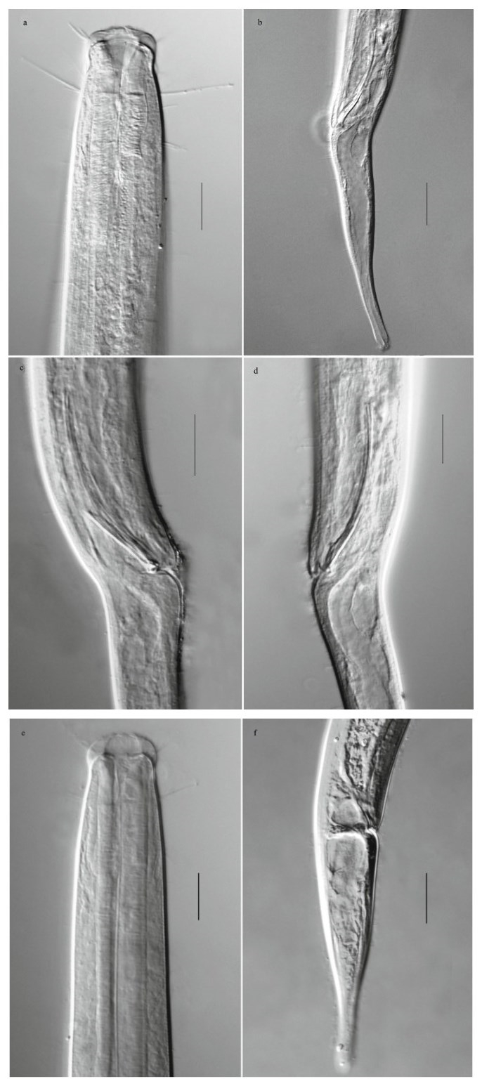
Males. Body cylindrical and gradually tapering towards tail end, 1 765–1 898 μm long and 46–52 μm wide at maximum body diameter. Head 27–28 μm wide. Cuticle with fi ne striations. A circle of eight long cervical setae, up to 28 μm, located just below the base of buccal cavity, about 26 μm from the anterior end.Numerous somatic setae scattered all over the body, about 7 μm long. Buccal cavity deep and large with hemispherical cheilostome and conical pharyngostome, Presence of a cuticular ring in the buccal portion. Anterior sensilla arranged in two circles. Six inner labial sensilla papilliform; six outer labial sensilla and four cephalic sensilla setiform and arranged in a circle, 12 μm and 6 μm long, respectively. Amphideal fovea not observed. Pharynx cylindrical and evenly sized to the end, about 20% of total body length. Pharyngointestinal junction with a large conical cardia, and surrounded by the intestine. Nerve ring at just before the mid of pharynx, about 40% of pharyngeal length from the anterior end. Excretory pore and ventral gland not seen. Tail conico-cylindrical, sharp narrowed of the body at the cloaca so that the tail slightly angled dorsally, 5.1–5.8 abd long, with distal third cylindrical and a swollen tip. Numerous caudal setae on both dorsal and ventral sides of the tail. Four caudal gland cells arranging in tandem, the anterior one extending beyond the level of the cloaca. The spinneret conspicuous, appearing as a cap covering the tip of tail. Three terminal setae 8 μm long.

|
| Fig.1 Paramonohystera weihaiensis sp. nov. a. lateral view of male anterior end; b. lateral view of male posterior end, showing spicules, gubernaculum and angled dorsally tail; c. lateral view of female head end, showing buccal cavity, cephalic setae and a circle of long cervical setae; d. lateral view of female tail end, showing caudal gland cells; e. entire view of female, showing ovary, oocytes and vulva. |

|
| Fig.2 Paramonohystera weihaiensis sp. nov. a. lateral view of male anterior end, showing buccal cavity, cephalic setae and a circle of long cervical setae; b. lateral view of male posterior end, showing spicules and angled dorsally tail; c. lateral view of male cloacal region, showing gubernaculum; d. lateral view of male cloacal region, showing spicule; e. lateral view of female anterior end, showing buccal cavity, cephalic setae and cervical setae; f. lateral view of female posterior end, showing tail and caudal gland cells (scale bar. a, c, d, e: 20 μm; b, f: 30 μm). |
Reproductive system diorchic, testes opposite and outstretched. Anterior testis to the left of intestine, posterior one to the right of intestine. Spicules slender and arcuate, 2.5 abd long, slightly cephalated proximally and tapered distally. Gubernaculum approximating as half long as spicule, plate-like, enlarged distal end with two teeth, without apophysis. No precloacal supplements observed.
Females. Similar to males, but slightly larger in body size, tail conico-cylindrical, not angled dorsally. A single long anterior outstretched ovary stretching to the pharyngeal base, situated to the left of the intestine. Ovary a wide tube, fi lled with oocytes. Uterus a short tube. Vagina straight. Spermatheca present. Vulva at the rear part of the body, about 70% of the body length from the anterior end.
3.6 Diff erential diagnosisParamonohystera weihaiensis sp. nov. is characterized by cervical region with a circle of long setae; tail sexual dimorphism, male tail angled dorsally with sharp narrowed of the body at the cloaca, female tail ordinary conico-cylindrical; amphideal fovea not observed; spicules slender, 2.5 abd long; gubernaculum plate, enlarged distal end with two teeth, without apophysis; precloacal supplements absent. Paramonohystera weihaiensis sp. nov. is close to Paramonohystera biforma Wieser, 1956 in general morphology. However, the described species has much slender body and larger "a" value (30.8–38.4 vs 18.3–22.2), shorter cephalic sensory setae ((11–12)+(5–6) μm vs (20–21)+(13–14) μm), absence of clear amphideal fovea (vs clear circle amphideal fovea), presence of a circle of long cervical setae (vs absence of circle of long cervical setae), spicules longer (2.5 vs 1.6–2 abd); gubernaculum odontoid distally (vs hooked distally). The new species is similar to Paramonhystera pellucida (Cobb, 1920) by the circle of long cervical setae but differs from the latter by the combination of its characteristics: tail sexual dimorphism, longer spicules (2.5 vs 2.0 abd), plate-like gubernaculum (vs tubular gubernaculum) and absence of clear amphideal fovea (vs presence of clear elliptical amphideal fovea). Besides, the new species differs from Paramonohystera sinica Yu & Xu, 2015 by larger body (1 765–2 004 μm vs 933–1 023), longer outer labial setae (11–12 vs 7–9 μm), relatively shorter spicules (2.5 vs 4–4.4 abd) and gubernaculum with distal tooth. The new species differs from Paramonohystera eurycephalus Huang & Wu, 2011 by the latter having clear round amphidial fovea (18–20 μm in diameter), longer spicules (3.1– 3.2 abd), tubular gubernaculum with a distal hook, fi ve to six precloacal supplements, absence of circle of long cervical setae.
4 DATA AVAILABILITY STATEMENTThe authors declare that the data supporting the fi ndings of this study are available within the article. The data will be available on request from the corresponding author.
5 ACKNOWLEDGEMENTThe authors are greatly thankful to Dr. HUANG Mian for his kind help in samples collection and slide making. We are sincerely grateful to three anonymous referees for providing valuable criticisms and improving on manuscript.
Bezerra T N, Decraemer W, Eisendle-Flöckner U, Hodda M, Holovachov O, Leduc D, Miljutin D, Mokievsky V, Peña Santiago R, Sharma J, Smol N, Tchesunov A, Venekey V, Zeng Z, Vanreusel A. 2018. Nemys: World Database of Nematodes. http://nemys.ugent.be. Accessed on 2018-12-01.
|
Chen G T, Vincx M. 2000. New and little known nematodes(Monhysteroida, Nematoda) from the strait of magellan and the beagle channel (Chile). Hydrobiologia, 429(1-3): 9-23.
DOI:10.1023/A:1003995005971 |
Cobb N A. 1920. One hundred new nemas (type species of 100 new genera). Contributions to a Science of Nematology, 9: 217-343.
|
Fonseca G, Bezerra T N. 2014. Order Monhysterida Filipjev, 1929 Handbook of Zoology. In:Schmidt-Rhaesa A ed.Handbook of Zoology. de Gruyter, Berlin. 435-465.
|
Gao Q, Huang Y. 2017. Oncholaimus zhangi sp. nov.(Oncholaimidae, Nematoda) from the intertidal zone of the East China Sea. Chinese Journal of Oceanology and Limnology, 35(5): 1 212-1 217.
DOI:10.1007/s00343-017-6114-5 |
Huang Y, Wu X Q. 2011. Two new free-living marine nematode species of Xyalidae (Monhysterida) from the Yellow Sea, China. Journal of Natural History, 45(9-10): 567-577.
DOI:10.1080/00222933.2010.534562 |
Huang Y, Zhang Y. 2014. Review of Pomponema Cobb(Nematoda:Cyatholaimidae) with description of a new species from China Sea. Cahiers de Biologie Marine, 55(2): 267-273.
|
Lorenzen S. 1977. Revision der xyalidae (freilebende Nematoden) auf der grundlage einer kritischen analyse von 56 arten aus nord-und ostsee. Veröffentlichungen Instituts für Meeresforschung in Bremerhaven, 16: 197-261.
|
Wieser W. 1956. Free-living marine nematodes. Ⅲ.Axonolaimoidea and Monhysteroidea. Acta Universitatis Lundensis (N. F. 2), 52(13): 1-115.
|
Xu Z, Huang Y. 2014. The research progress on free-living marine nematodes in China Sea. Journal of Liaocheng University (Natural Science Edition), 27(1): 55-60.
(in Chinese with English abstract) |
Yu T T, Xu K D. 2015. Two new nematodes, Pseudelzalia longiseta gen. nov., sp. nov. and Paramonohystera sinica sp. nov. (Monhysterida:Xyalidae), from sediment in the East China Sea. Journal of Natural History, 49(9-10): 509-526.
DOI:10.1080/00222933.2014.953224 |
 2019, Vol. 37


